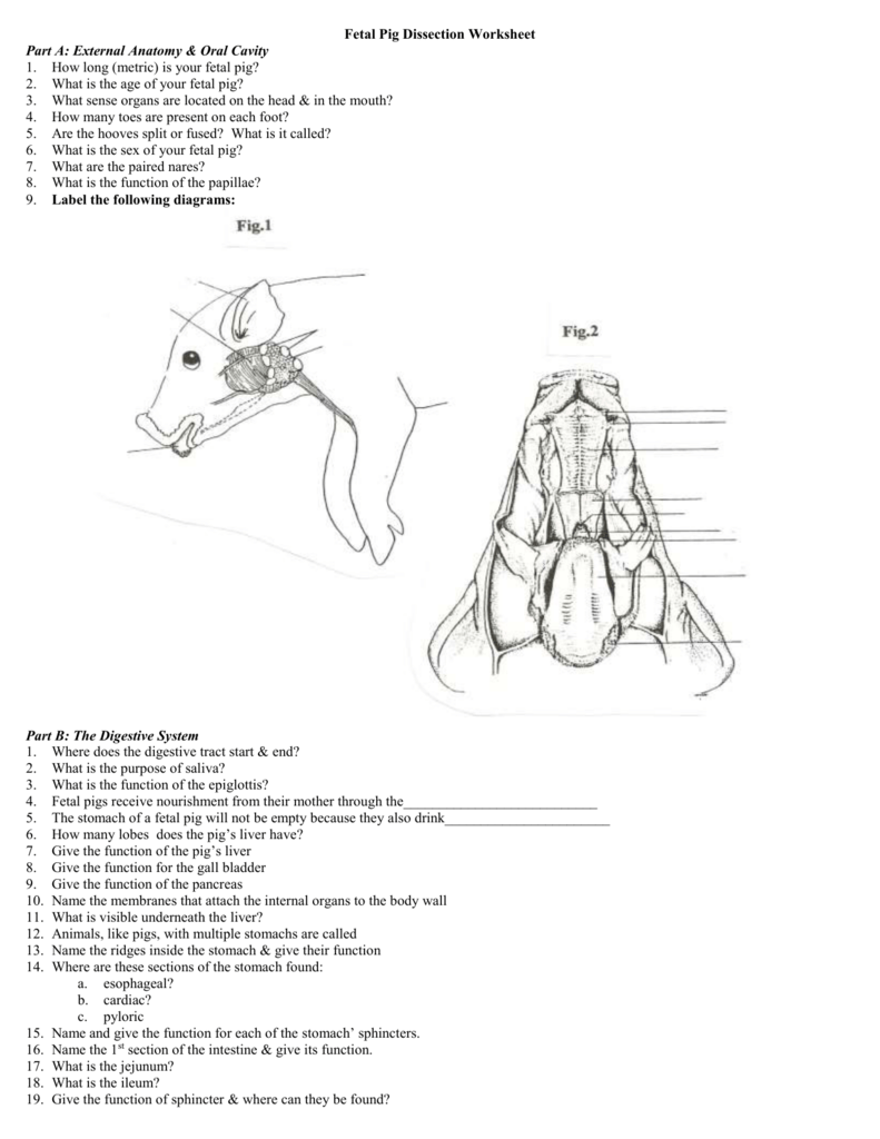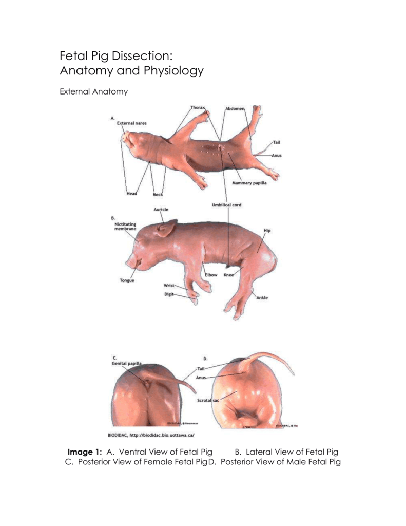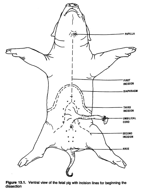

Diaphragm: a sheet of muscle and connective tissue that helps in breathing.See this diagram for the fetal pig heart, and the Wikipedia Heart article for some good diagrams of human heart anatomy. You may also see the right and left carotid arteries, which supply blood to the head. Note the aorta, where high-pressure blood leaves the heart on its way to the systemic circulation. The heart is surrounded by a pericardial sac. Look for these structures in the thoracic cavity: Note the many membranes lining the coelom and holding the organs in place. The thoracic cavity and the abdominal cavity are separated by the diaphragm. In mammals, the coelom is divided into two main cavities: the thoracic cavity, which contains the lungs, and the abdominal cavity, which contains the digestive system. Esophagus: carries food from mouth to stomach soft and muscular so it can move a food bolus by peristalsis.Trachea: the airway it's reinforced with rings of cartilage so it doesn't collapse.The thyroid secretes hormones that help regulate metabolism. Thyroid gland: another endocrine gland it’s a small bilobed structure just posterior to the larynx.It’s a large, spongy structure covering the ventral surface of the trachea and often extending into the thoracic cavity adjacent to the heart. Thymus gland: an endocrine (hormone-secreting) gland that helps regulate the immune system.If you cut it open, you can see the vocal cords inside. Larynx: an enlarged structure on the trachea.Locate and understand the functions of the following structures: Carefully separate the muscles to observe the underlying structures. Cut midline on the ventral surface of the neck to expose the underlying muscles. The more you cut things up, the harder it will be to figure out what you’re looking at. Once you open the body cavity, you will generally be able to separate the different organs by simply pulling them apart with your fingers, forceps, or a probe. Females: Look for the urogenital papilla, located just below the anus.īoth males and females have nipples, just as in humans.īegin your dissection in the neck region.The scrotal sac may be visible as a swelling just ventral to the anus, depending on the age of the fetus.The testes are still deep inside the body cavity they don't descend into the scrotal sac until later. Males: Urogenital opening is located near the umbilicus the penis is hidden inside.Here are some features you should look for: Determine the sex of your pigīefore you start dissecting, examine the outside of the pig and determine its sex.

The best online guide to fetal pig dissection is probably the Virtual Fetal Pig Dissection at Whitman College. Here's another specimen available in lab. On the photo below, I've labeled some structures that I might potentially ask you about on the lab exam. This would be a good one for the lab exam.

We have a dissected specimen in lab, preserved in a plastic case. Sagittally sliced pig sealed in plastic.Fetal pig that you can dissect with your group.Recognize the structures labeled on the pictures on this page or listed in bold in the text.

In addition, you should study the two pre-dissected specimens available in lab. In this lab you'll dissect a fetal pig to get a look at the anatomy of a mammal.


 0 kommentar(er)
